Were going to need a bigger boat…..and more time.

Greetings to you all

So you may be thinking that myself and Chris have died or forgotten about this little spot in the giant ocean that is the internet.

Well I can tell you that we are far from either of those in fact it’s far less rock and roll. During the time I was starting my blog I was courting the women who is I am pleased to say is now my fiancé. As you can imagine this has taken up a chunk of my time but also the PhD itself, which I am near to finishing (finally!) not to mention the paid work I have to do so I can live.

Chris has also had long list of things to sort out his end as well, the point is we have not much time to blog on the wonders of fossil fish.

This however is going to change soon..ish, ok roughly by next year I should be all done and I will have more time to blog, as I have really missed it.

Also I have another blog in the works so watch this space for those of you with a cryptozoology bend….

So until the next post, All the best.

He’s my cousin…well twice removed on my mother’s side

Well here at ancient anglers we like to be at the cutting edge of fish research and on that note I will now discuss a paper from last year…yeah. So last year a paper appeared in the journal Palaeontology, entitled “Origin of the White Shark Carcharodon (Lamniformes: Lamnidae) based on recalibration of the Upper Neogene Pisco Formation of Peru”. The reason this paper is so interesting is because it looks to solve the origins of one of the Earth’s marine apex predators the white shark (Carcharodon carcharias) (fig.1)

Image

Figure 1. The White shark (Carcharodon carcharias)

also known as the “great” white shark. However I suspect the reason it made the headlines is because of the words “white shark” and “Megalodon”, as we all know the general public has always be excited and fascinated my the idea of giant sharks  see films such as the critically acclaimed Jaws and the not critically acclaimed Mega shark vs. giant Octopus (fig. 2).

Image

Figure 2. The film you should all see!

So let’s get into it, what the paper aimed to do as already mentioned is work out the origin of the white shark however the paper also tries to constrain the age of key fossil baring horizons in the Pisco Formation using zircon Uranium–lead (U-Pb) dating and strontium-ratio analysis. So what does this involve? Well U-Pb dating is a radiometric dating method and while I won’t bore you with the ins and outs of dating, put simply as soon as a mineral forms in this case zircon the elements begin to decay and the length of time certain ones take can be used to date rocks to a rather accurate degree. Strontium-ratio analysis is used to investigate tooth enamel in archaeology as well as palaeontology, they use this to date their new species of Carcharodon to around (6-8 Ma).

So yes probably should mention the other purpose of the paper…the new species they describe C. hubbelli seen here (fig.3). Note that it’s only the teeth, jaws and some vertebrae, this is because sharks are not that hard…..well I would not say that to them in person, let me explain.

Image

Figure 3. Carcharodon hubbelli

Sharks belong to a class of animals called Chondrichthyans these critters skeletons are not made of bone instead a softer more flexible tissue called cartilage (we do have cartilage in our bodies located in the discs between our vertebrae and parts of ears and nose). The trouble is that in life cartilage is fantastic for an aquatic as it allows for a super flexible body, but rubbish if you want to be preserved in the fossil record as it’s softer nature means it breakdowns  quickly, which is why the majority of shark fossils are represented by teeth, the only “hard parts” on a shark.

The Pisco Formation is located on the coast of Peru and has some of the best Cenozoic marine fossils like elasmobranchs (sharks) teleost’s (the most common bony fish today) Turtles , shore birds and whales anywhere on the planet. So these are excellent deposits to find and try and understand the evolution of the White shark, so let’s try and understand the two schools of thought of how these sharks evolved. The first is that “megatoothed” sharks like Megalodon are part of the Lamnidae and that White sharks are close kin. However the second states that megatoothed sharks are in their own family, the Otodontidae within the Lamniformes (the Lamniformes being the apex ocean going predators of the shark world). What this means is that white sharks are not related to the megatoothed sharks at all.

This latter is the favoured view and the one certainly that the ancient anglers support, the issue with the first theory is that as with most shark work it’s based on teeth and sadly workers in the past have thought that the teeth of White sharks were similar to Megalodon, however it seems they did not take things like not knowing where in the mouth the tooth is located or whether the tooth has pedomorphic (features that are juvenile that an adult retains) characteristics.

So what does this mean for the overall evolution of the White shark, well think of the White shark as a beefed up Mako shark with broad teeth and when looking at them next to each other you can kind of see the similarities (fig. 4). What about the other sharks mentioned in the paper, well these turn out to be what are known as chrono-species, basically one species arose and replaced the other so in the case of the White shark and this paper it is proposed that the progression is Carcharodon hastalis-hubbelli carcharias (the White shark).

Image

Figure 4. (Left)Short fin Mako (Isurus oxyrinchus) (right) White Shark (Carcharodon carcharias)

Reference(s)

EHRET, D. J., MACFADDEN, B. J., JONES, D. S., DEVRIES, T. J., FOSTER, D. A. and SALAS-GISMONDI, R. (2012), Origin of the white shark Carcharodon (Lamniformes: Lamnidae) based on recalibration of the Upper Neogene Pisco Formation of Peru. Palaeontology, 55: 1139–1153. doi: 10.1111/j.1475-4983.2012.01201.x

All images taken from google images

Gone Fishin’ in the Jurassic

Is it really the end of January? Where does the time go?
It is high time I updated you on the Ancient Anglers’ antics. We’ve been fishing, sort of.
A couple of weeks ago on a bright, sunny Sunday, with a very low tide on the way, I picked up Luke (and Steve, of Mesozoic monsters fame) from Southampton station and we headed off to Dorset to visit the Jurassic coast. I’d love to tell you we were there on serious scientific business at the cutting edge of palaeontology, but the truth is we were indulging our love of fossil hunting. The plan was to visit Chapman’s Pool, a remote location about 3 miles East of Kimmeridge Bay.

Figure 1. Chapman's Pool from the cliff top.

Figure 1. Chapman’s Pool from the cliff top.

Figure 1 shows the view towards Chapman’s Pool from the top of the cliff. Unfortunately, I chose my cliff paths poorly, and the descent to the beach from where I took this photo would have been challenging to say the least! Rather than take the time to find the appropriate route to the beach, we decided to proceed to Kimmeridge Bay and park right next to the beach.
For those who don’t know this part of the world, the Dorset coast is a world heritage site with an almost complete succession of Jurassic strata exposed, getting younger as you move East. The area around Kimmeridge is late Jurassic and dates to between 155 and 150 million years ago. I won’t be speaking much of the geology in this post, but if you want to learn pretty much all there is to know about the geology and history of Kimmeridge (and many other areas) I highly recommend Dr. Ian West’s webpages.
http://www.southampton.ac.uk/~imw/Kimmeridge-Bay.htm
Dr. West has spent half a century exploring, cataloguing and studying most of the South coast and his webpages are filled with amazing photos and a wealth of information covering geology, palaeontology, petrology and local history.

Figure 2 looking East along the beach.

Figure 2 looking East along the beach.

Figure 2 shows the Kimmeridge Clay Formation looking East from Kimmeridge bay. Photo taken from the wave cut platforms which are exposed at low tide.
Since fossils were first discovered in the 19th Century at Kimmeridge, a huge diversity of prehistoric creatures has been found there. Dinosaurs, ichthyosaurs, plesiosaurs, pliosaurs and pterosaurs have all been found over the years. All very exciting and certainly of interest to us, but we were there for the fish!
Many genera of fish have been found at Kimmeridge including Aspidorhynchids, Coelacanths, Lepidotids, Pycnodonts, Thrissops and a host of other bony fish and sharks. Remains vary from isolated bones and teeth to exquisitely preserved partial and entire skeletons.
Figure 3 below shows one of the waterfalls that periodically adorn certain sections of cliff. One in particular was quite tricky to get past and is a cut-off point as the tide rises. The dark grey steps at the bottom are covered at high tide and this is a location where getting cut off is a real risk, so careful observations of tide and time is necessary. I include it here as I think it makes a nice photo!

Figure 3

Figure 3

Figure 4

Figure 4

Figure 4 was taken around one of the headlands looking West. Luke is waving in the distance. The entire area is underwater at high tide so we were trying to cover as much ground as possible in the available time. As well as searching for the more exciting (and predictably rare) fossils, there are some lovely ammonites preserved in the wave cut platforms. They cannot be collected as they are part of the platform, but some are very large and others well preserved. Here are a couple of examples we found (Figures 5 and 6.)

Figure 5

Figure 5

Figure 6

Figure 6

To be honest, as you search for the characteristic black and brown glint of bone, it is easy to become blasé about the sheer number of ammonites that are there. It’s easy to forget that these once living animals had to die, be buried and fossilised, and their remains survive for 150 million years, and then be exposed and then not be destroyed by erosion for us to be looking at them.
“Ammonite, ammonite, ammonite, big ammonite…” We stopped taking pictures of them quite quickly! (I’ve been on several field trips where finding even one ammonite would have made the day!) As I said earlier we were there for the fish, and they are relatively rare.
It is here that I wish I could insert a genuine “David Attenborough moment”, unveiling a spectacular discovery, as experienced by Steve a few months ago when he picked up a piece of rock in a quarry, telling us that this is exactly the sort of piece you might expect to find a tooth on, then turn it over to reveal a tooth!! I hope he blogged about that, it was a great moment, but I digress. As well as prospecting, we were there to investigate something I had found on a recent previous trip.

101_1016

Figure 5 shows a barnacle encrusted ammonite in the platform.
Figure 6 shows an impression of an ammonite, crushed flat.
Not very far East of the car park at Kimmeridge bay, about 7m from the cliff on a section of platform I found a collection of bones! It appeared to be the disarticulated remains of a partial skeleton of an individual animal! To say I was excited was an understatement. One of the reasons I love fossil hunting is the thrill of “treasure” hunting and to me that is the very definition of treasure.

101_0917

The problem I had was that it didn’t seem to fit anything I’d personally seen before. Some of the bones had a definite fishy sort of look (highly scientific, I know) but the straight bones were a confusing shape to me, and with the degree of weathering it was not immediately apparent what these bones are. So I thought it prudent to bring in some more learned fellows than I to have a look and try and identify what I’d found.
I’m pleased to report that they took the task very seriously. 

100_1037
After we stopped messing about, we quickly eliminated most of the possibilities, and concluded it had to be a fish of some sort, but the section of 7 vertebrae, much smaller than the other bones, must have come from another fish, preserved at the same time. On top of that, it definitely wasn’t a coelacanth, or (unsurprisingly) any of the Palaeozoic fishes that Luke studies. From the size of the bones we knew it had to be a fairly large fish, as no small fish has straight bones that large, but we had no further ideas at the time.

100_1036

101_0966.

I include a couple of close up shots to highlight both the find itself and the difficulty in finding diagnostic bones in certain fossil assemblages. More photos are available on request.
As a significant collection of bones found in Kimmeridge bay is rare, it is important that these finds are reported. I had a fairly big clue that I was not the first person to find these bones. A 30 x 30cm (approx.) section had been buzz sawed out of the platform right next to my find, and there is only one person legally able to do this. I raised the subject when I was taking some photos of the coelacanths he found that I am studying.
Turns out my “awesome” exciting find is not museum worthy. It is a fish, it is rare, but it is not well enough preserved and not enough of it is preserved to excavate given the expense of so doing. As can be seen from the photos it is very weathered.
It has been identified as a large Pycnodont.

These are a group of fish that had dentition well adapted to crushing shells and came in a variety of sizes and body forms. A lot of them were laterally compressed and oval or circular in outline. This one was clearly a big one!


Whilst I am disappointed I didn’t find something museum worthy, I am pleased to note something that not everyone gets to see. In a few tides’ time (1-5 years at most) this will be gone forever. I encourage all of you that have an opportunity to visit the area to have a look, take some snaps. Here is a shot of the cliff directly opposite the bones.

101_0970

The two cut out bits at the bottom of the cliff are quite distinctive.

All in all, it was a very enjoyable day. We prospected as far as we dared whilst the tide was low, saw some nice fossils and began and ended our trip looking at the best fossils I’ve ever found in the Jurassic. And it was a fish, so technically we were fishing, and we caught one!

That’s about all from me for this post. I’ll be in touch soon.

Chris


From Estonia…..with fish?

It has to be said that sometimes international research collaboration is underrated and if I’m being honest I was not sure what to expect from my trip to Estonia. I will return to what I got from it at the end of the post.

Image

Figure 1. Location of Estonia

So first off where is Estonia and why did I go out there, Estonia is just below Finland on the map (fig.1)

Image

Figure 2. Dr Tiiu Märss

and the capital Tallinn is located on the coast of the Baltic sea. I went there for a very simple reason….knowledge, you see I have been at this PhD for a year and bit now, and all my knowledge about the fish in my bone bed is self-taught. The reason for this is that although the University of Portsmouth has experts in both vertebrates and the Silurian neither have worked on Agnatha, so this is why I have had to teach myself.  Now luckily for me, my supervisors know all sorts of researchers including Dr. Tiiu Märss (fig.2)

who for a number few years has worked on Palaeozoic fish so she was going to give me first-hand knowledge on these groups so that I could identify the disarticulated material in my bone bed. So the way I’m going to do this post is tackle each day and what I got up to.

Monday  

BEEP BEEP BEEP, oh god its 3am I didn’t even know this time existed! So two trains and a 2 hour and 45 minute flight and I find myself in Estonian….Tallinn to be exact and once I have located my bag I headed for the door, where I was met by Tiiu (holding up a sign saying “LUKE” very rock & roll).

After a bus ride across the city I reached my home for the next four days the Academic hostel, which was very good and I would highly recommend it, any who so after a spot oflunch we made it to the department which as you can see in (fig.3) is located next to a

Image

Figure 3. no it’s not Narina

forest! The first thing we did was chat in her office about my project and I showed her my samples I brought from the UK.  After this I was shown to my work area (fig.4) which was all very swish and I started going through her UK material.

Image

Figure 4. The office and my work place for the four days

Tuesday

So day 2 begins with where I left off the previous day, sat at the binocular microscope (fig.5) and as mentioned looking through the loose samples that Tiiu has collected over the years and this was useful as I could get a sense of what certain scales look like and ask Tiiu questions about the material. I felt very lucky to be looking at all these different localities that I have read about in papers and places I know are historically important like some of the Scottish sites. Another interesting point was the variety of preservation in the denticles, browns, blacks and whites colours, and of all qualities. While looking, every so often I would stop and draw a denticle of interest and identifying them, I had a great many slides to through at least 60 so this was going to take some time. It was also great to know that the denticles in my sample which I had already identified as Paralogania ludlowensis was correct as Tiiu confirmed this (this is not surprising as this is expected to be the most common genera in my bed…but time will tell).

Image

Figure 5. My set up with Microscopes and note pad oh yeah and the samples (far left)

Wednesday

Now Wednesday was an interesting one as I was able to look at a large number (some 20 drawers) of articulated agnatha and gnathostomes (that’s animals in this case fish with jaws). Before that I did finish off looking through those disarticulated specimens….partly because I had not finished looking through them as there were so many and well partly because I had over slept on the Tuesday oops!

So I descended into the basement of the institute (the natural home of the palaeontologist) where their fossil collection is located, then as with the disarticulated material I sat down with a microscope and slowly went through each one stopping to draw ones of interest. Now one thing some of you readers may not realised is that there is a very good reason why they call people like myself a micro-palaeontologists and that’s because you spend most of your time strapped to a microscope of one type or another and this certainly was the case in Estonia. In term of the material it was a mixed bag some of the specimens were to be honest a little grotty while others were insanely beautiful, of particular note was the Canadian artic specimens which I was told by Tiiu were the “rubbish” bits that Wilson did not want when they were collecting in the 1990’s. So I know at this point I would normally saying here “see fig.1” I will be 100% honest folks I did have a chance to take pictures at this point, however this photo taken in Tiiu office (fig. 6) shows an articulated specimen of Phlebolepis elegans and the coolest part of this is that I have seen this actual specimen in the flesh….or denticles I guess.

Image

Figure 6. A photo of a photo of Phlebolepis elegans

After that there was a quick coffee and cake break, the coffee was great and served in their own mug! (fig.7) How cool is that, the poppy seed and chocolate bun was lovely as well. Back to work and I was looking at a “small” section of a mass mortality bed from one of the Islands of Estonian (Saaremaa in particular) and to describe it, well think of those Green River Formation slabs covered in fish it’s like that except with Phlebolepis elegans in the Silurian!

Image

Figure 7. Yes the institute has thir own mugs!

Thursday

The last day before flying home on the Friday was mostly made up of three things, looking through the Estonian material like I did with the UK material, which was excellent, seeing the differences between the two areas….oh I should probably explain, ok so back in the Silurian Earths continents looked rather different (fig.8.) The UK was known as Avalonia a series of small islands…not Scotland though as it was part of America….until it slammed into Wales and England while Estonia was part of the continent known as Baltica. The second thing was getting a load of papers……some in Russian…..still the pictures are nice the translation will come later I think. Finally, what I like to think as a bit of a highlight is some thin section photography I did with my thin sections on her microscope and camera (that’s the one on the left in fig.5) and as you can see in (fig.9) the results were excellent with all manner of details seen.

Image

Figure 8. Map showing the Silurian paleocontinents of Avalonia and Baltica

So what are my final thoughts about my first international research trip, well overall excellent it was really good chatting to a fellow thelodont researcher and gives me a real sense of what I need to do and how to do it. I recommend to my fellow researcher if you can go out to another part of the world and learn from others and to see other stuff to very much go and do it, as it’s so very useful. Also a final note I am stood in down town Tallinn and waiting for a bus to the airport when who should I bump into but my friend Charlotte from my local college back in my home town…..it truly is a small world.

Luke.

Image

Figure 9. A denticle from cut through horizontally, you can see the dark borwn in the centre which is the pulp cavity and the thin brown lines which are the dentine tubules and the ornimentation on the sides of the denticle

Well there you are I hope you have enjoyed this first year of Ancient anglers we are very sorry we have not posted more but we hope this will change next year. To all our readers, followers and causal browsers Merry Christmas and a happy new year.

from the Ancient Anglers

If you have liked the blog this year why not like our Facebook page and be first to get the latest updates and fishy news, click here to go there and hit like, thanks.

References

Figure 2 is from http://www.gi.ee/index.php?page=30&staff_id=36 and figure 1 and figure 8 comes from Google images.

Better late than never – the other ancient angler speaks!

Hello all, Chris here.
I think it is about time for me to join the blog and start posting. This post will be quite short and aims to introduce myself to you all, and outline what I am researching.
Like Luke, I am also a PhD student at Portsmouth University, and for my sins I aim to be a fish worker too, or Palaeoichthyologist to give us our posh title! However, despite graduating with Luke in 2011, my personal circumstances meant that my PhD research has begun a year later so I am a little behind the work that Luke has done to date.
My research concerns a group of fish known as the Actinistians, the Coelacanth family. These are a fascinating group of fish that until 1938 were thought to have gone extinct at the KT boundary along with the (non-avian) Dinosaurs, 65 million years ago.
They first appeared in the fossil record in the Early Devonian – c.400 MYA, and we still have 2 species swimming in our oceans today. They survived both the Permo-Triassic major extinction and the KT (Cretaceous/Tertiary) extinction, left no (yet found) evidence of their existence for 65 million years, and were then discovered alive in the 1930s (fig.1).

Figure 1. latimeria chalumnae (The living coelacanth)

I will be studying some Late Jurassic Coelacanth fossils, found by Steve Etches, the world renowned collector of Kimmeridgian (Late Jurassic from mostly Kimmeridge Bay) fossils, and aim to ascertain their relationship with other Coelacanths. It is possible that these fossils represent new species, new to science.

Future posts will detail the family, what we know so far and will include some pictures!
In addition to the Coelacanth related posts, I am a keen fossil hunter, so I will indulge myself with the occasional post to show off my finds and explain what I have found and where it came from.
Yours piscatorially
Chris

References

Images taken from Google images

So what are doing with your time exactly Mr Hauser?

Hello Luke here, here it is then my first post on fish related to my research, sorry it’s so late folks but it’s been quite hectic. But before I introduce the cast of characters I will just mention what my PhD is on. I am looking at the Downton bone bed from the welsh borders; some of you may be wondering “Downton bone bed? Well I have heard or the very famous Ludlow bone bed”. This is kind of the point of my research, when you look at the Ludlow bone (fig.1) you can see this grey lithology and all those black grains…their fossils and in particular fish fragments. This is also true of the Downton bone bed however if you were to see it you would not clearly see the fossils, this is because all the fossil grains are of a similar colour to the lithology (a tan/brown colour) and this is why it has been missed for full investigation for the last 150+ years. So the next few sections for you delectation are some brief summaries of the types of fossil fish that I will come across, of course one the most exciting aspects of my PhD is the potential of finding new things, now this does not necessarily mean new species but possibly fish that are not known from this part of the world or at this time or who knows what. So without further ado here are the cast of characters I will become familiar with over the next possible 6 years and hopefully you guys will learn to love them, these odd bunch of early fish.

Image

Figure 1 Ludlow Bone Bed

Condonts

So these little jawless beauties appear in the Cambrian and make it all the way to the end Triassic! That is very impressive when you consider a lot of these fish don’t make it past the P/T mass extinction let alone animals getting past the Devonian. While on the subject of “animals” let’s clear things up about condonts and conodont animals, now condonts have been know about since 1856 when Christian Pander a Latvian embryologist and palaeontologist first identified them but, they were not the whole animal. They are calcium phosphate (or to be all fancy like calcium carbonate fluroapatite) microfossils known as conodont elements (fig.2)

Image

Figure 2. Conodont elements: (a, b) coniform elements, (c, d) ramiform elements, (e, f) pectiniform blade elements, (g, h) pectiniform platform elements, (i) bedding-plane assemblage.

which would have been found in the mouth of the animal (fig.3) Now these elements can be categorised by shape into different types protoconodonts, paraconodonts and euconodonts and due to taxa like Ozarkodina whose apparatus has been found complete and articulated we know how they looked in the living animal (fig.4).

So what about the living animals well the conodont animal or as some

Image

Figure 3. (A) Dorsal view of the reconstructed, closed apparatus of Novispathodus.(B) Orientation of the apparatus within the conodont’s head.

researchers are suggesting they should be called Conodontophora (conodont bearers), either works for me. These were eel like fish (fig.5) ranging from a less than a centimetre to those gigantic Ordovician monsters of the Soom shale which were 10’s of centimetres. Now it is at this point I should explain some of the many controversies with conodonts, so first off; what are these chaps (and chapettes) eating, well the main idea is that these were filter feeders using that complex apparatus in the mouth to filter out plankton from the water column. However in more recent years researchers have found that the elements have microwear patterns and this tells us that these guys are grasping prey so they are more likely swift little hunters.

Image

Figure 4 (a) natural assemblage of conodonts from the Carboniferous of Illinois (×24); and (b) the conodont animal from the Carboniferous Granton Shrimp Bed, Edinburgh, Scotland, with the head at lefthand end (×1.5).

Other blog posts in the works for these critters are; Conodonts: are they vertebrates? (yes and here’s why) and were conodonts venomous?

Thelodonts

Ah thelodonts or to translate their name ‘nipple tooth’, these jawless fish (agnatha) ranging from a few centimetres to meter in length and make up the bulk of the bone bed fauna so far seen. Now I apologise if I wax lyrical about these guys but in terms of my project I have come across hundreds of these guys but like 4 conodont elements so you can understand if these get a tad more press but worry not fans of conodonts they will get their own post.

Image

Figure 5. Reconstructions of Conodontophorans in life

So let’s get into them, they appear in the Middle or Late Ordovician and sadly bit farewell and join the choir invisible in the Late Devonian. We find their fossils can be found all over the world and we can find them as complete articulated specimens but the majority of their fossils (and certainly this is the case with my material) they are found as isolated micro fossils (fig. 6). I should at this point explain what thelodonts are; they are fish with an endoskeleton of cartilage and then there skin is covered in dermal denticles (fig.7) in a slightly analogous way with sharks. The denticles, as the dent part of the word suggests are related to teeth…that is for want of a better word they are teeth in the skin (hence the dermal bit). In the fossil record they usually “rock” (no pun intended) up as isolated denticles in sediment most famously in the Late Silurian bone beds but occasionally as entire articulated specimens found in places like Canada, Scotland and Scandinavia with all their denticles in place. While on the subject of places we find their remains add to the list Russia, Australia, South East Asia, Europe and finally North and South America. They are a monophyletic group although one group the Furcacaudiformes from Canada are morphologically very different and there is debate on their position in thelodonti and represent a new order of thelodonts (Märss, 2006a).

Image

Figure 6. thelodont fish in life

Overall thelodont affinities remain somewhat unclear, it has been suggested that they are the sister taxa to the osteostracans and could also be close to chondrichthyes but the way thelodonts have their micromeric squamations (scales) is different from both. Ultimately when it comes to where these fish fit one must remember there we are talking about a group of animals mostly known from there isolated dermal denticles in fact in most decent phylogenetic analyses only contains24 to 25 taxa because that’s how many we have articulated. Now that’s not to say that that makes it invalid I am unsure how many characters were generated but 24 taxa is quite a small selection but does once again highlight the challenges one faces when dealing with the fossil record.

On a final foot note for now diet wise these chaps are probably eating plankton, algae and rubbish (well deposit and suspension feeding on sediment) in fact the Furcacaudiformes with their deep bodies and large eyes were suggested to be predators. Right well not top of the line predators but still taking on small prey items however it is more likely the large eyes were used to help select only the tastiest bits of detritus….also helps in spotting predators, like giant sea scorpions.

Image

Figure 7. thelodont denticles Loganellia scotia

Acanthodians

The final group of fish that I will be looking at and discussing here are the “spiny sharks” or acanthodians (fig. 8) named in 1844 by Agassiz (for more information about Agassiz’s life and work click here). This group gets its name from the Greek word akanthos which means spine and it’s well placed as they are the only fish that processed spines on all their fins. They appeared in the Silurian which is handy for me and disappeared when coprolite hit the fan with end Permian extinction. However one of the most notably features of the acanthodians are jaws…yes these are some of the first jawed vertebrates but at the time of my research (roughly 420 ma) they are not the dominant fish fauna it’s still the agnathans. There found in the fossil record like thelodonts both as, in my case isolated fragments of denticles or as articulated specimens and they are found in all corners of the globe. In terms of there relationships with other early fish we rule out early sharks (although the spiny sharks  name came from morphological similarities) placoderms and early actinopterygians, but of the jawless ancestors only thelodonts come close to a similar micromeric squamation but it is unlikely the two are that closely related. It now seems that they share a lot of specialized characters with osteichthyans compared to other groups, some of the characters are: similar shaped brain case, nature of the gill filaments (hemibranchs) along the gill arch and the presence of brachiostegal rays. Although (Davis et al 2012), found that they are somewhat split between the osteichthyans and the chondrichthyans, so once again this shows that with dealing with fish this far back getting a complete resolution of their origins can be difficult.

Image

Figure 8. Example of an acanthodian Climatius reticulatus, Illustrator Arthur Weasley

There are three subgroups of acanthodians the first is the Climatiiformies which were I guess the “standard” acanthodian although were more heavily armoured than the other two. The next it is the Ishnacanthiformes which had robust jaws and big teeth and assumed to be the predators of the group and finally the Acanthodiformes which were the most successful of the groups and despite developing jaws became filter feeders (turns out have jaws is not just handy for grabbing food items). The final bit of classification is the scale morphology there being two types, one is the Acathodes type which is a crown made of true dentine and a thick acellular bone base and Nostolepis type that has a dentine crown penetrated by vascular canals and a base of cellular bone.

Others

Now there have also been chondrichthyan scales found in bone beds of this age in the welsh borders but apart from saying “there are sharky things in them there hills” I can’t expand really, I also may come across material from other types of agnathans and as soon as the thesis or paper is accepted I will be blogging about it, I also plan to do more in depth posts on these groups but all in good time.

So yes there we are that’s the sort of fish I will be working on hopefully Chris will tell you about his fish soon so until next time (which will be a special post) take care.

References

Davis, S. P.; Finarelli, J. A.; Coates, M. I. 2012. “Acanthodes and shark-like conditions in the last common ancestor of modern gnathostomes”. Nature 486 (7402): 247.

Janvier. P, 1996. Early Vertebrates, Clarendon press. Oxford. Pp.393

Long. J.A, 2010. The rise of fishes: 500 million years of evolution, Johns Hopkins University Press. pp.304

Märss, T. Turner, S. & Karatajūtė-Talimaa, V. 2007. Handbook of paleoichthyology Volume 1B “Agnatha” II Thelodonti, pp.141.

Märss, T., 2006a. Exoskeleton ultrasculpture of the early agnathans and fishes. – Journal of vertebrate paleontology. 26(2), p.235-252.

All taken from Google images apart from figure 4 taken from http://www.blackwellpublishing.com/paleobiology/figure.asp?chap=16&fig=Fig16-5&img=c16f005.

The SVPCA 2012 spectacular!

Yes yet again apologies for the delay from the last blog post but we have been busy bee…fish? Anyway yes Chris is preparing to start his PhD and I have been rushing around and for the last two weeks so here is the belated SVPCA blog post oh and for those wondering what does SVPCA mean (symposium of vertebrate palaeontology and comparative anatomy).

Ok so let us start with a very brief overview of the week, the conference this year was held in the beautiful city of Oxford one of the Holy Grail’s of early British palaeontology. The venue was the fantastic Oxford University Natural History Museum which in terms of a venue for this sort of thing is just the tops. So it was a weeklong lecture series and how this works is thus day one: fish, days two and three: reptiles and days four and five: mammals (terms and conditions may vary please ask in store for full details). So course for this blog please understand it’s just fish talks and posters discussed here, however there were loads of talks I would love to discuss, sadly I cannot though do check out the links at the bottom of this post for other blogs discussing other talks. The other side of SVPCA is the social this is not purely a bunch of academics getting ratted….honest; no it’s more of a networking session, a chance for fellow workers to meet and or catch up. I alone have some exciting projects in the pipe line thanks to these casual chats over free coffee and wine (incidentally my love for wine increases directly in relation to its price….so at SVPCA I LOVED the wine). So that is the extra bits of SVPCA done, now on to the meat and potatoes of the event.

So the way I’m going to tackle these talks and posters is somewhat alphabetically according to the author however may not be in the order I heard them etc. So first up was a talk entitled:

An overview of the hybodont record of the Cameros Basin (northwest of Iberian Range, Spain), Dr. David Didier Bermúdez-Rochas presented this thought provoking talk on the diversity of hybodont (fig.1)

Figure 1 Hybodont reconstruction

sharks in the Cameros basin of Spain which to be honest according to the talk was rubbish but that could be due to the continental sediments. The basin’s previous taxa were referred to Asteracanthus ornatissimus, Hybodus polybrion and Hybodus sp. so yeah not the vast assemblage you might expect. However surface prospecting in a new locality showed a much larger assemblage Lonchidion (which is represented by the highest number of elements), Lissodus, Parvodus, Hybodus, Planohybodus and Egertonodus, that’s more like it and these provide a window into a freshwater community of hybodonts being the dominant freshwater sharks (and we thought we had problems with just bull sharks). This new data provides evidence that the faunas of northwest Spain now have similarities with other regions of Spain as well as the classic Wealden localities of Britain. It was suggested by colleagues that this flush of new taxa may be due misidentification in not accounting for the change in teeth within parts of the mouth. Now certainly this is true of in the jaw reconstruction of sharks like Carcharocles megalodon as workers did not account for the dental pattern, however this is not a fair argument for this talk as that is more to do with size than morphology so I am confident with the identification of these sharks.

3D textural analysis of microwear and trophic ecology of placodermsfirst of all congrats to Mr Laurent Darras whose talk this was who has just passed his viva with minor corrections so now he is Dr. Laurent Darras. Now to the talk given not by Laurent but by his supervisor Dr. Mark Purnell, the talk discussed the work Darras has done on morphospace on fossil aquatic gnathostomes (jawed vertebrates). The idea of morphospace analysis is that by simply plotting points on a jaw or a whole fossil you can create shapes these shapes can be plotted and basically you get pretty graphs which can suggest what ecology these animals have. The problem he points out is that sometimes what the morphospace suggests is not always what the animal actually feeds on so you need independent evidence to either to confirm the morphospace data or not. In the discussion the placoderms, that’s fish like Dunckleosteus(fig.2)

Figure 2 Dunkleosteus

were used as a model to test if microwear analysis could be used to test the idea of microwear being a good bit of independent analysis. The other questions this could answer like ecomorphology, ontogenetic shift in diet and predation driven macroevolution events. He has been able to prove that microwear analysis can be used to test and constrain specific hypotheses of the diet and trophic diversity in all kinds of fish. I remember at progressive palaeontology he discussed this with Pycnodont fish…..via the medium of lord of the rings….you had to be there!

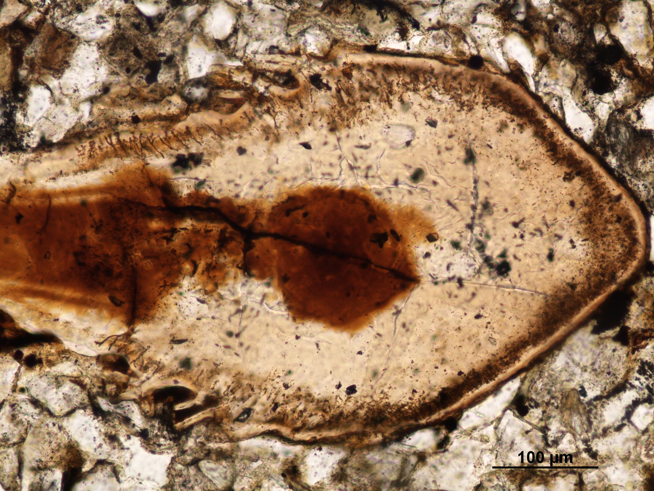
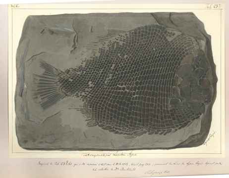
Evolution of bone repair via invasive growth of dentine in a 380 million year old fish, this talk was present by Dr. Zerina Johanson who in my very short career I have already had the pleasure of working with, So this talk centres around a specimen of Psammolepis (fig.3)

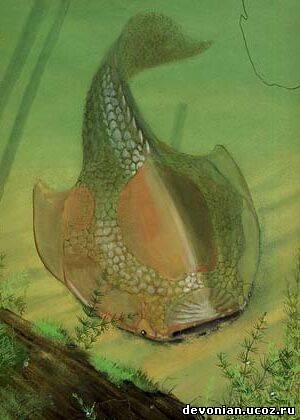
Figure 3 Psammolepis

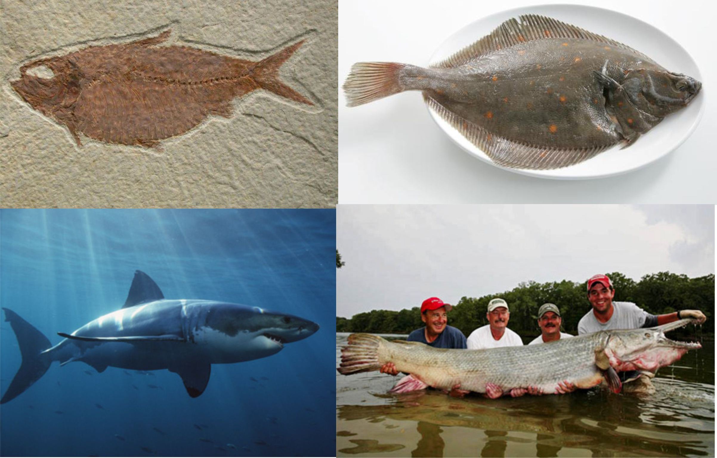
a Heterostracan (one of the many groups of jawless fish I will talk about in the next post). Now this specimen is interesting because of the damage on the dorsal surface of the fish not necessarily because of the predator-prey interactions, but as the title suggests from the point of view of the repaired damage. The skeleton of these jawless fish are dominated by dermal done in a form known as aspidin which is a precursor to bone as we would know it, on the surface however there are many dentine tubules (fig.4). The deep injury to the Psammolepis is cool because it shows evidence of healing this in itself is not surprising (an animal healing injury ruddy Nora!) no what is interesting is that the fish has used dentine to heal the wound, literally flooding it. The bone (aspidin) appears to have not been involved at all like the fat kid never picked for sports. The dentine is produced and laid down in a chaotic manner by ondontoblasts which are found in the tubercle pulp cavities as well as in the surrounding flask shaped crypts in the bone. Ok now at this point I could talk about the idea in the talk about stem cells and pore canal systems etc, but what I personally found amazing is that sand grains managed to get included in the healed wound this inclusion is not so shocking as you can see from the image of Psammolepis it’s flatted body is indicative of a modern flat fish and I can easily imagine these chaps partially buried in the sediment of the seafloor and this is how the sand ended up in the wound and being covered in dentine.

Figure 4 The simplifed structure of Psammolepis demal plate

Beating the bends: The spare ribs of Big Meg now this talk was given by the ever so entertaining Dr. Jeff Liston, and concerned the large amount of bones belonging to the giant Jurassic pachycormid Leedsichthys (fig.5) (and yes you guessed it were going to do a post on these guys too). Now without getting to bogged down these are BIG fish some estimates go up to 53 feet (Liston, 2005) and being fish they have a truck load (literality!) but a lot are broken and fragmented and ultimately what you end up with is a load of ‘rib-shaped bones’…not that helpful. So what Jeff has tried to do is to reverse engineer the bones so that we can see where they may been on the body as the idea being that a bone reflects the stresses and pressures that is was under during life so if you normalise the bone you can have a better idea where is goes. So here is the maths: normalised bone curvature = X/2L x 100…….ok got that good simply what he found is that this by using some Argentinean specimens where we know the accurate position of the bones and there curvature, he could plot the Leedsichthys elements into three size clusters which showed that many came from the anterior. A slight curve ball here is the fact, and I was unaware of this is that the older the pachycormid in age (which sometimes meant the larger they were) the less the skeleton is ossified which makes sense when you think about the sheer size of these leviathans (in weight saving etc).

Figure 5 Leedsichthys & Liopleurodon

New specimens of Symmoriidae sharks from the Carboniferous limestone of the Derbyshire Peak District the shark themed talk was presented by the lovely Kelly Richards who is a PhD student at Cambridge University and was an introduction of her project, what she has done and what she hopes to do. We learn that fossils are full of ‘bucket taxa’, synonymies, inadequate diagnostic descriptions etc and this is very true in the Palaeozoic shark family Symmoriidae. Within the talk new material from two horizons in Derbyshire were mentioned one Stethacanthus altonensis(fig. 6)

Figure 6 Stethacanthus

which until this point has only been known from Bear gulch in Montana and is represented by quite substantial material which will help clear things up and Akmonistion zangerli(fig.7) known previously from Bearsden deposits Scotland. Again this taxon is represented by substantial material which should help build a better picture of the palaeobiology and paleogeography of the Symmoriidae. Over all being a fellow PhD I always enjoy these talks, that is ones explaining the aims of the projects and being able to see the projected direction of the PhD so kudos to Kelly there.

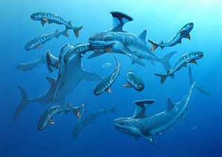
Figure 7 Akmonistion

Lazarus Shark taxa in the Triassic of Australia Dr. Sue Turner came with another shark talk (because sharks are cool!) and this as the name suggests is on a ‘Lazarus taxa’, now some of you reading this may be wondering what Lazarus taxa means well Lazarus was a brother in the Bible who came back to life (no not a zombie!) and this case it’s when a taxa disappears from the fossil record only to reappear later on either in the fossil record or in the modern day. The coelacanth is a prime example of a Lazarus taxa, but back to the matter at hand, xenacanthiform sharks what about them, well Sue told us how new material have been found and this with material already known and described in 1908 by Arthur Smith Woodward. The talk raised an interesting point that the fossil record of xenacanths (fig.8)

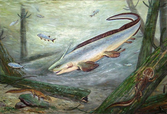
Figure 8 Xenacanthus

is well known in the Lower Carboniferous of Queensland but none are known from the later  Palaeozoic expect for these new and old specimens from the Triassic. So how did these sharks get through the Permo-Triassic extinction and was Australia a refuge for the sharks, hopefully further investigation of this new material will in time answer these questions

Phew well there we are….oh wait hold on forgot the posters so without further of do:

New chondrichthyan-like scales from the Lower Silurian of Mongolia it was nice to see this poster by Plamen Andreev, being a fellow Silurian micro vertebrate worker, the take home message I got from this poster is that a new chondrichthyan has been found this however is based on scales rather than a complete fossil animal. I was intrigued by the use of micro-CT and it’s something I would be tempted to possibly try this on my own scale material in the future.

New details about the Ordovician jawless fish Sacabambaspis janvieri Marco Castiello who I have chatted to many times before SVPCA (check out his blog here) was there with his beautiful poster featuring artwork from the very talented Stefano Broccoli. There poster was very exciting as the work concerned the agnathan (jawless) fish Sacabambaspis(fig.9)

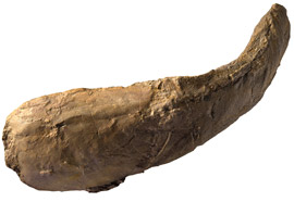
Figure 9 fossi of Sacabambaspis

one of the first well preserved vertebrates whose remains have been found across the world. It always amazes me that even something like this well known fish…..honestly it’s well known, is not exactly what we thought and the study carried out suggests the oral paltes are composed by some small platelets that are similar to lepidotrichia as well a number of other features.

Evolution and development of a morphological innovation: The pufferfish beak (Tetraodontiformes; Teleostei)this poster by Gareth Fraser was frankly a brilliant piece of work and as the title suggests looked at the beaks of pufferfish (fig.10)

Figure 10 Pufferfish

using normarsky optics, micro-CT and optical projection tomography to show that although the pufferfish beak looks like an innovative structure it’s not made from a new genetic blueprint, oh no it’s just evolution tinkering with the general osteichthyan dentition, truly the beauty sometimes is in the detail.

So that’s about it folks for this post, oh I did have a poster there but I don’t like sounding my own trumpet but I think it went down well…ish. Hope you enjoyed this review hopefully soon I will post up my PhD fish related goodness until then stay safe and say no to drugs.


Blogs with SVPCA posts

Mesozoic monsters

Ichthyosaurs: a day in the life…..

gimpasauria

Paleostories

References

Liston, JJ (2005). Homologies amongst the fragments: searching for synapomorphies in shattered skulls. In: Poyato-Ariza FJ (ed) Fourth International Meeting on Mesozoic Fishes – Systematics, Homology and Nomenclature, Extended Abstracts. Servicio de Publicaciones de la Universidad Autónoma de Madrid/UAM Ediciones, Madrid, pp 141–145.

All images from Google images

Switzerland, not just famous for knives and Toblerone

Luke here with a new post, I promise that I will soon deliver a more academic post about the sorts of fossil fish I study but first I thought I would use this post to discuss a very special gentleman. I am not sure how many of your good selves know this, but us fish workers owe a huge amount to this man (fig 1).

Figure. 1 Louis Agassiz

Jean Louis Rodolphe Agassiz or Louis Agassiz for short is frankly the father of Paleoichthyology (the study of fossil fish). Born in May 1807, Môtier-en-Vuly, Switzerland, the son of a protestant Pastor. He was schooled at home from an early age, but after finishing his elementary education at Lausanne, he decided to study medicine which he did at the universities of Zürich, Heidelberg and Munich. While at these institutions he made good use of their facilities and became interested in natural history, particularly botany. It’s at this point some of you may be wondering “where is he going with this”…..hold on I’m getting there.  Now up to this point he had not shown any interest in fish but this is how he fell into them as it were, these two chaps J.B Spix and C.F.P von Martius had popped back from a successful trip to Brazil and had collected all manner of things including fish from the Amazon. Spix kicked the bucket long before he got to work on the history of these fish so Martius selected the fresh out of uni Agassiz to work on them instead. By 1829 the work was published as Agassiz threw himself into the work, this was followed by a study of the history of the fishes in the Lake of Neuchâtel. In 1830 he then released a prospectus on History of the Freshwater Fishes of Central Europe which was completed in 1842; 10 years earlier in 1832 he was appointed the professor of natural history at the University of Neuchâtel. Now a self proclaimed fish addict (well not shouting from the tops of mountains….then again he was Swiss!) it was not long before fossil fish attracted his gaze. Fossil fish were well known from the slates of Glarus and the limestones of Monte Bolca but no one had actually scientifically studied them until Agassiz rocked up (sorry for the pun).

Figure. 2 Example of the Dinkle watercolours

It was this work that would bring him his worldwide fame with his enthusiasm and work effect he churned out 5 volumes of his Recherches sur les poissons fossils between 1833 and 1842, illustrated beautifully by Joseph Dinkel (fig 2), on his travels to different museums (something our Chris is going to understand all to soon!) he met the great Georges Cuvier (French anatomist and all round swell guy) who helped and encouraged him greatly. So while Agassiz was working on these fossils he realised that it was necessary to develop a new classification scheme as there were no soft tissues preserved, instead fin rays, teeth and scales made up the bulk of the collections. To this end he came up  with the four groups based on scales and other dermal appendages viz. Ganoids, Placoids, Cycloids and Ctenoids (fig.3), sadly today this form of classification does not really work on a modern classification scheme as pointed out by A.S. Woodward. As this work continued it was clear that (as PhD students know all too well) he was running out of wonga, luckily aid was at hand in the form of the Earl of Ellesmere who loved Dinkel’s images so much he brought all 1290 of them and gave them to the Geological Society of London (we will come back to this later). The same society gave him the Wollaston medal and made him a foreign member for his services to ichthyology.

Figure 3. a) Cycloid scale (Pike), b) Ctenoid scale (Perch), c Placoid scale (Thornback), d Placoid scale of Rhina and e Ganoid scales (Palaeoniscus

On his first visit to England in 1834, Hugh Miller and other geologists brought to light the remarkable fishes of the Old Red Sandstone of the northeast of Scotland. The odd fish they found like Pterichthys, the Coccosteus were naturally of intense interest to Agassiz, from which he produed a special monograph in 1844-45: Monographie des poissons fossiles du Vieux Grès Rouge, ou Système Dévonien (Old Red Sandstone) des Iles Britanniques et de Russie. He went on to do so much more in both geology and zoology and travelled from North and South America a number of times before his death in 1873 at Cambridge, MA. He is buried at Auburn and has a monument at his grave of an erratic boulder from the moraine of the glacier of Aar (this cost a pretty penny I can tell you!) from where he did some of his best known geological work.

So why tell you lovely people all this, well remember I said about those images that were given to the Geological society of London, good because they need your help in preserving them. They are old and knackered and these beautiful watercolours need our help to restore them and allow them to be digitised so all can enjoy them forever. So why not sponsor-a-fish (click the link) just £20 will help protect and keep forever one watercolour out of some 2,000 in the collection each person who donates will get there name on a roll of honour (which is very nice) but more importantly you will be succouring this important bit of scientific history for everyone to enjoy. From July of this year they have made some £6000 which is excellent but their target is £20,000 and would be great if we can help them do this.

So I hope you have enjoyed reading about this man’s impressive life he is not the only key player in Paleoichthyology and I’m sure we will post up biographies on other important fish workers in the future. Until next time stay safe and don’t talk to strange people unless they are you teacher or lecturer then you don’t have a choice.

Text refernces

for a more in depth look at Agassiz’s life click here

Image references

All images taken from google images

The Difficult first post

So hello and welcome to the blog, as it says in the about section the reason for myself and Chris creating this blog is that if you look out there on the internets you’ll find all manner of blogs on fossil organisms from dinosaurs to mammals and everything else if you go on Tetrapod zoology!

But you will find very few on fish, yes that’s right those extremely common vertebrates (representing some 27,977 of the 54,711 known vertebrates, that’s over one-half!!!) represented by some 515 families both extinct and extant. They are possibly the most morphologically diverse group of vertebrates; I mean think about it look at a Knightia alta, a Plaice (Pleuronectes platessa), a white shark (Carcharodon carcharias) and a Alligator gar (Atractosteus spatula) figure.1 (I haven’t even mentioned seahorses!), they are all extreme shapes, it seems a little unkind to say “well fish are fish shaped” too such a fantastically diverse group. They also inhabit a wide range of habitats from the polar oceans to the muddy banks of mangrove forests. Not to mention exhibiting a huge range of bizarre and fascinating behaviours.

Fig. 1 (Top left) Knightia, (Bottom left) White shark, (Top right) Plaice, (Bottom right) Alligator gar.

Now some of you cladistics fans will be shouting “but what are fish?” and “Fish don’t exist” and yes it is true that fish are a paraphyletic group this is because they are “basal” their characters can be found in a wide range of vertebrates making it hard to identify them as an actual group, “fish” is a colloquial term, however I think we’re going to have to write a blog post about this topic all on its own.

Now if we take the wonders of extant fish, we can only imagine what fossil fish over the last 500 ma must have been like, and this is the plan of the ancient anglers to inform and entertain about fossil and at times extant fish topics that interest us, important papers and cool things about fish.

Anyway that’s about it from the this first post thanks for staying for the ride hope this gave you a flavour for what is to come from myself and Chris, the posts may be a bit sporadic for the first few months as we find the time to write these but hopefully we should have at least a couple a month.

References

Nelson. J. S, 2006. World of fishes, John Wiley & sons inc, Hoboken, New Jersey. pp.601

Long. J.A, 2010. The rise of fishes: 500 million years of evolution, Johns Hopkins University Press. pp.304

All images are from Google images