Yes yet again apologies for the delay from the last blog post but we have been busy bee…fish? Anyway yes Chris is preparing to start his PhD and I have been rushing around and for the last two weeks so here is the belated SVPCA blog post oh and for those wondering what does SVPCA mean (symposium of vertebrate palaeontology and comparative anatomy).
Ok so let us start with a very brief overview of the week, the conference this year was held in the beautiful city of Oxford one of the Holy Grail’s of early British palaeontology. The venue was the fantastic Oxford University Natural History Museum which in terms of a venue for this sort of thing is just the tops. So it was a weeklong lecture series and how this works is thus day one: fish, days two and three: reptiles and days four and five: mammals (terms and conditions may vary please ask in store for full details). So course for this blog please understand it’s just fish talks and posters discussed here, however there were loads of talks I would love to discuss, sadly I cannot though do check out the links at the bottom of this post for other blogs discussing other talks. The other side of SVPCA is the social this is not purely a bunch of academics getting ratted….honest; no it’s more of a networking session, a chance for fellow workers to meet and or catch up. I alone have some exciting projects in the pipe line thanks to these casual chats over free coffee and wine (incidentally my love for wine increases directly in relation to its price….so at SVPCA I LOVED the wine). So that is the extra bits of SVPCA done, now on to the meat and potatoes of the event.
So the way I’m going to tackle these talks and posters is somewhat alphabetically according to the author however may not be in the order I heard them etc. So first up was a talk entitled:
An overview of the hybodont record of the Cameros Basin (northwest of Iberian Range, Spain), Dr. David Didier Bermúdez-Rochas presented this thought provoking talk on the diversity of hybodont (fig.1)
sharks in the Cameros basin of Spain which to be honest according to the talk was rubbish but that could be due to the continental sediments. The basin’s previous taxa were referred to Asteracanthus ornatissimus, Hybodus polybrion and Hybodus sp. so yeah not the vast assemblage you might expect. However surface prospecting in a new locality showed a much larger assemblage Lonchidion (which is represented by the highest number of elements), Lissodus, Parvodus, Hybodus, Planohybodus and Egertonodus, that’s more like it and these provide a window into a freshwater community of hybodonts being the dominant freshwater sharks (and we thought we had problems with just bull sharks). This new data provides evidence that the faunas of northwest Spain now have similarities with other regions of Spain as well as the classic Wealden localities of Britain. It was suggested by colleagues that this flush of new taxa may be due misidentification in not accounting for the change in teeth within parts of the mouth. Now certainly this is true of in the jaw reconstruction of sharks like Carcharocles megalodon as workers did not account for the dental pattern, however this is not a fair argument for this talk as that is more to do with size than morphology so I am confident with the identification of these sharks.
3D textural analysis of microwear and trophic ecology of placodermsfirst of all congrats to Mr Laurent Darras whose talk this was who has just passed his viva with minor corrections so now he is Dr. Laurent Darras. Now to the talk given not by Laurent but by his supervisor Dr. Mark Purnell, the talk discussed the work Darras has done on morphospace on fossil aquatic gnathostomes (jawed vertebrates). The idea of morphospace analysis is that by simply plotting points on a jaw or a whole fossil you can create shapes these shapes can be plotted and basically you get pretty graphs which can suggest what ecology these animals have. The problem he points out is that sometimes what the morphospace suggests is not always what the animal actually feeds on so you need independent evidence to either to confirm the morphospace data or not. In the discussion the placoderms, that’s fish like Dunckleosteus(fig.2)
were used as a model to test if microwear analysis could be used to test the idea of microwear being a good bit of independent analysis. The other questions this could answer like ecomorphology, ontogenetic shift in diet and predation driven macroevolution events. He has been able to prove that microwear analysis can be used to test and constrain specific hypotheses of the diet and trophic diversity in all kinds of fish. I remember at progressive palaeontology he discussed this with Pycnodont fish…..via the medium of lord of the rings….you had to be there!
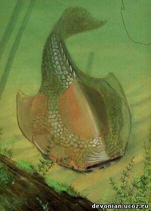
Evolution of bone repair via invasive growth of dentine in a 380 million year old fish, this talk was present by Dr. Zerina Johanson who in my very short career I have already had the pleasure of working with, So this talk centres around a specimen of Psammolepis (fig.3)
a Heterostracan (one of the many groups of jawless fish I will talk about in the next post). Now this specimen is interesting because of the damage on the dorsal surface of the fish not necessarily because of the predator-prey interactions, but as the title suggests from the point of view of the repaired damage. The skeleton of these jawless fish are dominated by dermal done in a form known as aspidin which is a precursor to bone as we would know it, on the surface however there are many dentine tubules (fig.4). The deep injury to the Psammolepis is cool because it shows evidence of healing this in itself is not surprising (an animal healing injury ruddy Nora!) no what is interesting is that the fish has used dentine to heal the wound, literally flooding it. The bone (aspidin) appears to have not been involved at all like the fat kid never picked for sports. The dentine is produced and laid down in a chaotic manner by ondontoblasts which are found in the tubercle pulp cavities as well as in the surrounding flask shaped crypts in the bone. Ok now at this point I could talk about the idea in the talk about stem cells and pore canal systems etc, but what I personally found amazing is that sand grains managed to get included in the healed wound this inclusion is not so shocking as you can see from the image of Psammolepis it’s flatted body is indicative of a modern flat fish and I can easily imagine these chaps partially buried in the sediment of the seafloor and this is how the sand ended up in the wound and being covered in dentine.
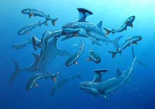
Beating the bends: The spare ribs of Big Meg now this talk was given by the ever so entertaining Dr. Jeff Liston, and concerned the large amount of bones belonging to the giant Jurassic pachycormid Leedsichthys (fig.5) (and yes you guessed it were going to do a post on these guys too). Now without getting to bogged down these are BIG fish some estimates go up to 53 feet (Liston, 2005) and being fish they have a truck load (literality!) but a lot are broken and fragmented and ultimately what you end up with is a load of ‘rib-shaped bones’…not that helpful. So what Jeff has tried to do is to reverse engineer the bones so that we can see where they may been on the body as the idea being that a bone reflects the stresses and pressures that is was under during life so if you normalise the bone you can have a better idea where is goes. So here is the maths: normalised bone curvature = X/2L x 100…….ok got that good simply what he found is that this by using some Argentinean specimens where we know the accurate position of the bones and there curvature, he could plot the Leedsichthys elements into three size clusters which showed that many came from the anterior. A slight curve ball here is the fact, and I was unaware of this is that the older the pachycormid in age (which sometimes meant the larger they were) the less the skeleton is ossified which makes sense when you think about the sheer size of these leviathans (in weight saving etc).
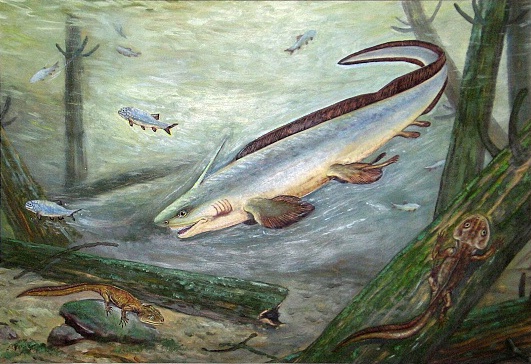
New specimens of Symmoriidae sharks from the Carboniferous limestone of the Derbyshire Peak District the shark themed talk was presented by the lovely Kelly Richards who is a PhD student at Cambridge University and was an introduction of her project, what she has done and what she hopes to do. We learn that fossils are full of ‘bucket taxa’, synonymies, inadequate diagnostic descriptions etc and this is very true in the Palaeozoic shark family Symmoriidae. Within the talk new material from two horizons in Derbyshire were mentioned one Stethacanthus altonensis(fig. 6)
which until this point has only been known from Bear gulch in Montana and is represented by quite substantial material which will help clear things up and Akmonistion zangerli(fig.7) known previously from Bearsden deposits Scotland. Again this taxon is represented by substantial material which should help build a better picture of the palaeobiology and paleogeography of the Symmoriidae. Over all being a fellow PhD I always enjoy these talks, that is ones explaining the aims of the projects and being able to see the projected direction of the PhD so kudos to Kelly there.
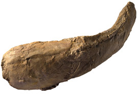
Lazarus Shark taxa in the Triassic of Australia Dr. Sue Turner came with another shark talk (because sharks are cool!) and this as the name suggests is on a ‘Lazarus taxa’, now some of you reading this may be wondering what Lazarus taxa means well Lazarus was a brother in the Bible who came back to life (no not a zombie!) and this case it’s when a taxa disappears from the fossil record only to reappear later on either in the fossil record or in the modern day. The coelacanth is a prime example of a Lazarus taxa, but back to the matter at hand, xenacanthiform sharks what about them, well Sue told us how new material have been found and this with material already known and described in 1908 by Arthur Smith Woodward. The talk raised an interesting point that the fossil record of xenacanths (fig.8)
is well known in the Lower Carboniferous of Queensland but none are known from the later Palaeozoic expect for these new and old specimens from the Triassic. So how did these sharks get through the Permo-Triassic extinction and was Australia a refuge for the sharks, hopefully further investigation of this new material will in time answer these questions
Phew well there we are….oh wait hold on forgot the posters so without further of do:
New chondrichthyan-like scales from the Lower Silurian of Mongolia it was nice to see this poster by Plamen Andreev, being a fellow Silurian micro vertebrate worker, the take home message I got from this poster is that a new chondrichthyan has been found this however is based on scales rather than a complete fossil animal. I was intrigued by the use of micro-CT and it’s something I would be tempted to possibly try this on my own scale material in the future.
New details about the Ordovician jawless fish Sacabambaspis janvieri Marco Castiello who I have chatted to many times before SVPCA (check out his blog here) was there with his beautiful poster featuring artwork from the very talented Stefano Broccoli. There poster was very exciting as the work concerned the agnathan (jawless) fish Sacabambaspis(fig.9)
one of the first well preserved vertebrates whose remains have been found across the world. It always amazes me that even something like this well known fish…..honestly it’s well known, is not exactly what we thought and the study carried out suggests the oral paltes are composed by some small platelets that are similar to lepidotrichia as well a number of other features.
Evolution and development of a morphological innovation: The pufferfish beak (Tetraodontiformes; Teleostei)this poster by Gareth Fraser was frankly a brilliant piece of work and as the title suggests looked at the beaks of pufferfish (fig.10)
using normarsky optics, micro-CT and optical projection tomography to show that although the pufferfish beak looks like an innovative structure it’s not made from a new genetic blueprint, oh no it’s just evolution tinkering with the general osteichthyan dentition, truly the beauty sometimes is in the detail.
So that’s about it folks for this post, oh I did have a poster there but I don’t like sounding my own trumpet but I think it went down well…ish. Hope you enjoyed this review hopefully soon I will post up my PhD fish related goodness until then stay safe and say no to drugs.
Blogs with SVPCA posts
Ichthyosaurs: a day in the life…..
References
Liston, JJ (2005). Homologies amongst the fragments: searching for synapomorphies in shattered skulls. In: Poyato-Ariza FJ (ed) Fourth International Meeting on Mesozoic Fishes – Systematics, Homology and Nomenclature, Extended Abstracts. Servicio de Publicaciones de la Universidad Autónoma de Madrid/UAM Ediciones, Madrid, pp 141–145.
All images from Google images


