It has to be said that sometimes international research collaboration is underrated and if I’m being honest I was not sure what to expect from my trip to Estonia. I will return to what I got from it at the end of the post.

Figure 1. Location of Estonia
So first off where is Estonia and why did I go out there, Estonia is just below Finland on the map (fig.1)

Figure 2. Dr Tiiu Märss
and the capital Tallinn is located on the coast of the Baltic sea. I went there for a very simple reason….knowledge, you see I have been at this PhD for a year and bit now, and all my knowledge about the fish in my bone bed is self-taught. The reason for this is that although the University of Portsmouth has experts in both vertebrates and the Silurian neither have worked on Agnatha, so this is why I have had to teach myself. Now luckily for me, my supervisors know all sorts of researchers including Dr. Tiiu Märss (fig.2)
who for a number few years has worked on Palaeozoic fish so she was going to give me first-hand knowledge on these groups so that I could identify the disarticulated material in my bone bed. So the way I’m going to do this post is tackle each day and what I got up to.
Monday
BEEP BEEP BEEP, oh god its 3am I didn’t even know this time existed! So two trains and a 2 hour and 45 minute flight and I find myself in Estonian….Tallinn to be exact and once I have located my bag I headed for the door, where I was met by Tiiu (holding up a sign saying “LUKE” very rock & roll).
After a bus ride across the city I reached my home for the next four days the Academic hostel, which was very good and I would highly recommend it, any who so after a spot oflunch we made it to the department which as you can see in (fig.3) is located next to a
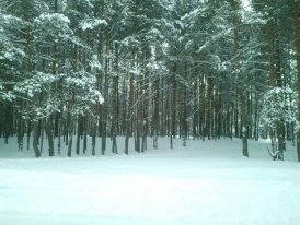
Figure 3. no it’s not Narina
forest! The first thing we did was chat in her office about my project and I showed her my samples I brought from the UK. After this I was shown to my work area (fig.4) which was all very swish and I started going through her UK material.
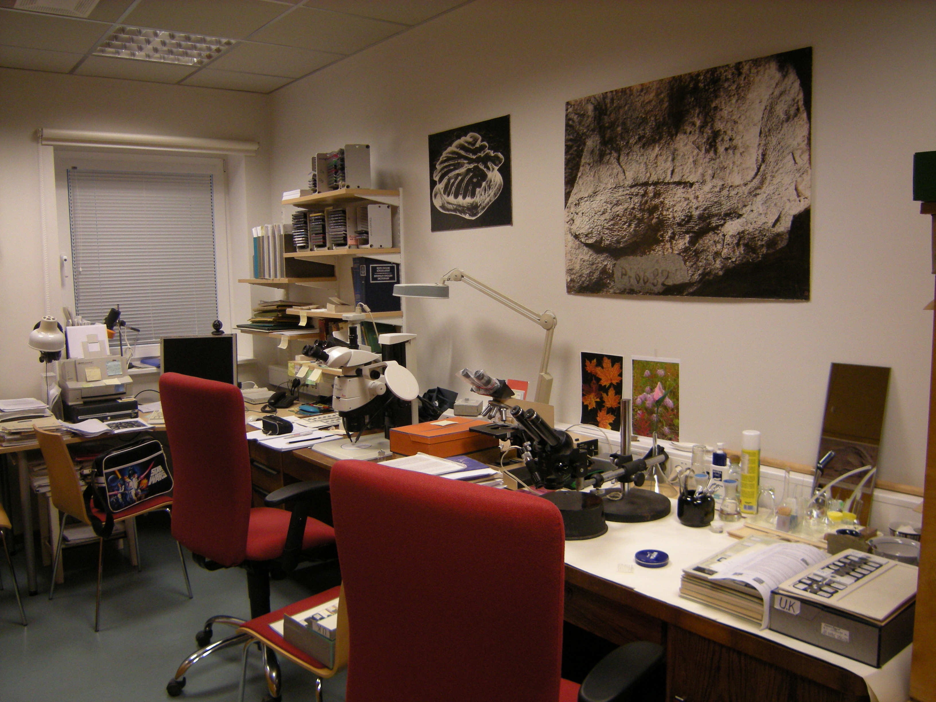
Figure 4. The office and my work place for the four days
Tuesday
So day 2 begins with where I left off the previous day, sat at the binocular microscope (fig.5) and as mentioned looking through the loose samples that Tiiu has collected over the years and this was useful as I could get a sense of what certain scales look like and ask Tiiu questions about the material. I felt very lucky to be looking at all these different localities that I have read about in papers and places I know are historically important like some of the Scottish sites. Another interesting point was the variety of preservation in the denticles, browns, blacks and whites colours, and of all qualities. While looking, every so often I would stop and draw a denticle of interest and identifying them, I had a great many slides to through at least 60 so this was going to take some time. It was also great to know that the denticles in my sample which I had already identified as Paralogania ludlowensis was correct as Tiiu confirmed this (this is not surprising as this is expected to be the most common genera in my bed…but time will tell).

Figure 5. My set up with Microscopes and note pad oh yeah and the samples (far left)
Wednesday
Now Wednesday was an interesting one as I was able to look at a large number (some 20 drawers) of articulated agnatha and gnathostomes (that’s animals in this case fish with jaws). Before that I did finish off looking through those disarticulated specimens….partly because I had not finished looking through them as there were so many and well partly because I had over slept on the Tuesday oops!
So I descended into the basement of the institute (the natural home of the palaeontologist) where their fossil collection is located, then as with the disarticulated material I sat down with a microscope and slowly went through each one stopping to draw ones of interest. Now one thing some of you readers may not realised is that there is a very good reason why they call people like myself a micro-palaeontologists and that’s because you spend most of your time strapped to a microscope of one type or another and this certainly was the case in Estonia. In term of the material it was a mixed bag some of the specimens were to be honest a little grotty while others were insanely beautiful, of particular note was the Canadian artic specimens which I was told by Tiiu were the “rubbish” bits that Wilson did not want when they were collecting in the 1990’s. So I know at this point I would normally saying here “see fig.1” I will be 100% honest folks I did have a chance to take pictures at this point, however this photo taken in Tiiu office (fig. 6) shows an articulated specimen of Phlebolepis elegans and the coolest part of this is that I have seen this actual specimen in the flesh….or denticles I guess.

Figure 6. A photo of a photo of Phlebolepis elegans
After that there was a quick coffee and cake break, the coffee was great and served in their own mug! (fig.7) How cool is that, the poppy seed and chocolate bun was lovely as well. Back to work and I was looking at a “small” section of a mass mortality bed from one of the Islands of Estonian (Saaremaa in particular) and to describe it, well think of those Green River Formation slabs covered in fish it’s like that except with Phlebolepis elegans in the Silurian!
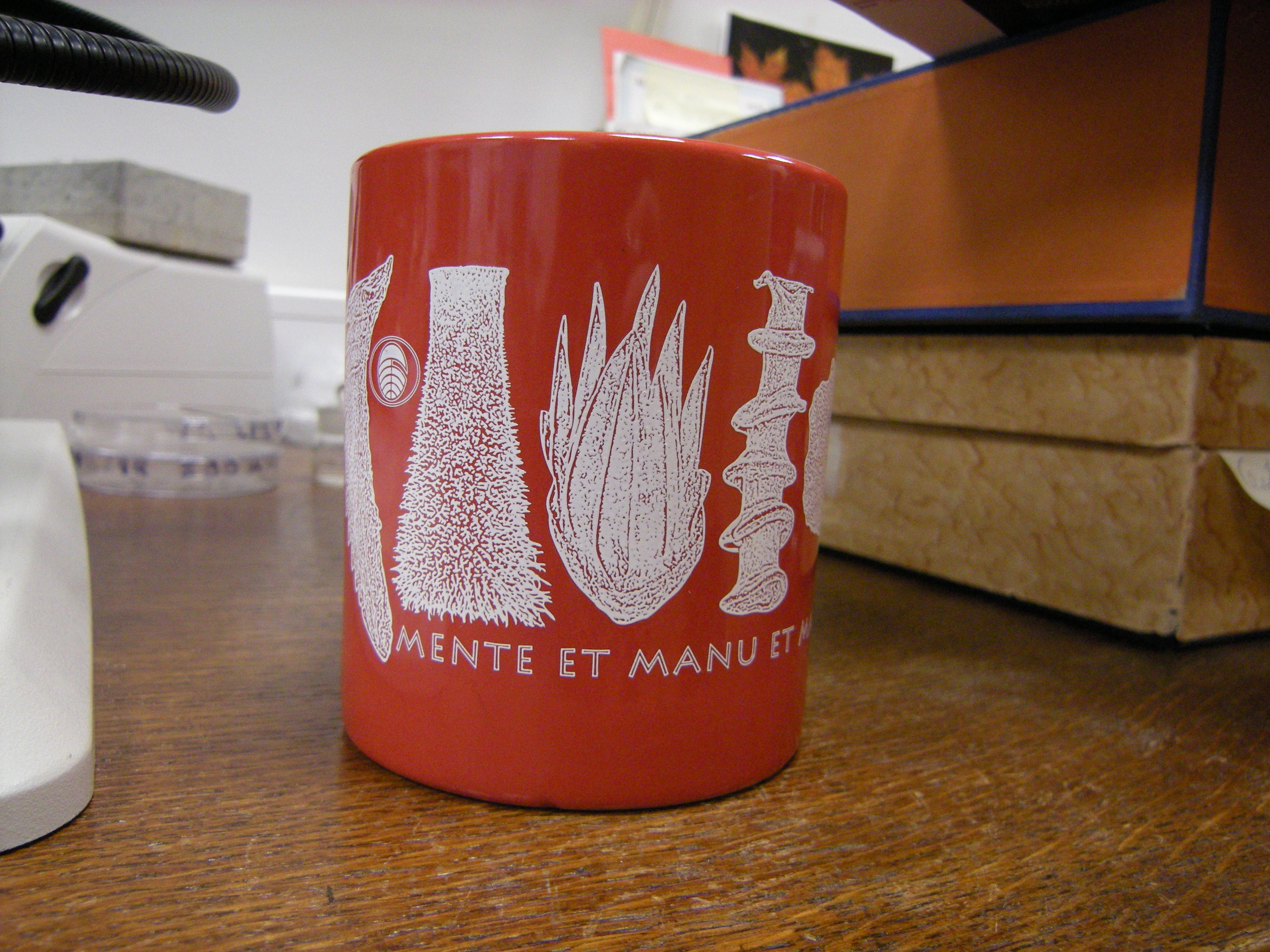
Figure 7. Yes the institute has thir own mugs!
Thursday
The last day before flying home on the Friday was mostly made up of three things, looking through the Estonian material like I did with the UK material, which was excellent, seeing the differences between the two areas….oh I should probably explain, ok so back in the Silurian Earths continents looked rather different (fig.8.) The UK was known as Avalonia a series of small islands…not Scotland though as it was part of America….until it slammed into Wales and England while Estonia was part of the continent known as Baltica. The second thing was getting a load of papers……some in Russian…..still the pictures are nice the translation will come later I think. Finally, what I like to think as a bit of a highlight is some thin section photography I did with my thin sections on her microscope and camera (that’s the one on the left in fig.5) and as you can see in (fig.9) the results were excellent with all manner of details seen.
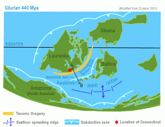
Figure 8. Map showing the Silurian paleocontinents of Avalonia and Baltica
So what are my final thoughts about my first international research trip, well overall excellent it was really good chatting to a fellow thelodont researcher and gives me a real sense of what I need to do and how to do it. I recommend to my fellow researcher if you can go out to another part of the world and learn from others and to see other stuff to very much go and do it, as it’s so very useful. Also a final note I am stood in down town Tallinn and waiting for a bus to the airport when who should I bump into but my friend Charlotte from my local college back in my home town…..it truly is a small world.
Luke.
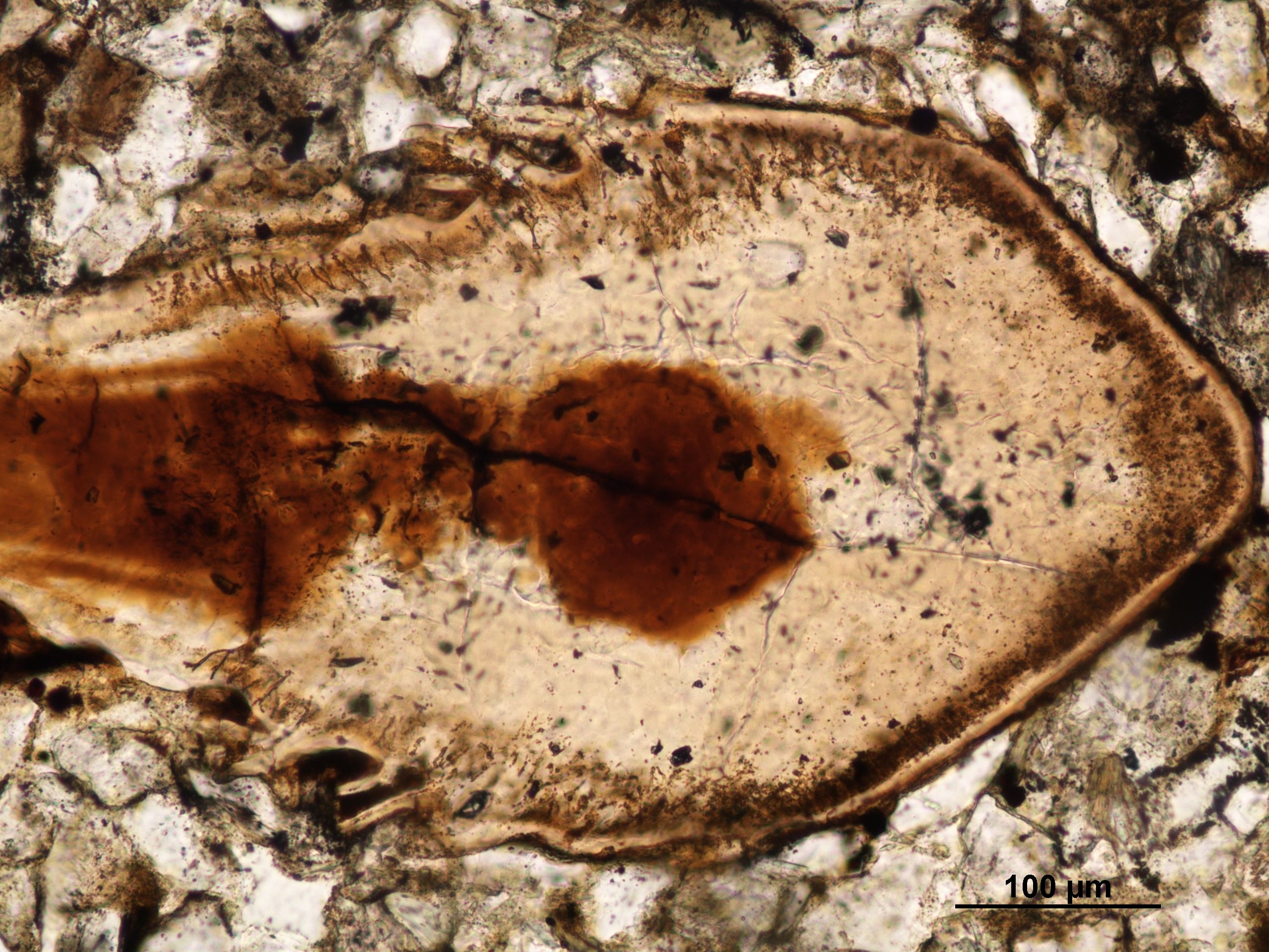
Figure 9. A denticle from cut through horizontally, you can see the dark borwn in the centre which is the pulp cavity and the thin brown lines which are the dentine tubules and the ornimentation on the sides of the denticle
Well there you are I hope you have enjoyed this first year of Ancient anglers we are very sorry we have not posted more but we hope this will change next year. To all our readers, followers and causal browsers Merry Christmas and a happy new year.
from the Ancient Anglers
If you have liked the blog this year why not like our Facebook page and be first to get the latest updates and fishy news, click here to go there and hit like, thanks.
References
Figure 2 is from http://www.gi.ee/index.php?page=30&staff_id=36 and figure 1 and figure 8 comes from Google images.
